
DISSECTION OF PALAEMON (PRAWN)
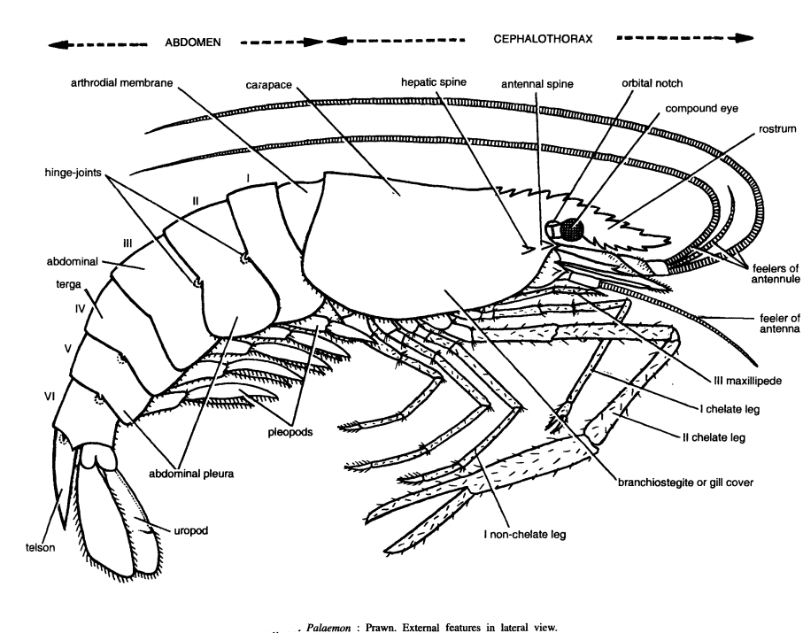
External features of Palaemon (Prawn)
It is fresh-water prawn, which forms a highly palatable dish. Prawn is the most favourite and well-liked animal by the students.
- Total length : 25 to 35 cm.
- Shape: Spindle-shaped, elongated and bilaterally symmetrical.
- Colour: Pale blue or greenish it becomes orange-pink on preservation in formalin or on boiling.
- Exoskeleton: It forms a ring around the segment. On dorsal side, the exoskeleton is in the form of a convex shield, called as tergum; the downward projection of tergum is called as pleuron and ventral shield is called as sternum. The hardened pieces or sclerites and segments of the appendages are joined together by soft articulating membranes, called as arthrodial membranes.
- Divisions of the body: The body of the animal is composed of nineteen appendage-bearing segments. It is regionated into anterior (i) cephalothorax and posterior (ii) abdomen. Abdomen is slightly flexed.
- Cephalothorax : It is large, rigid, unjointed and cylindrical in shape and is formed by 5 appendage-bearing head segments and 8-appendage-bearing thoracic segments. Exoskeleton of cephalothorax is formed by carapace, which is dorsally and anteriorly produced into a serrated rostrum. On the sides, carapace hangs freely and covers gills and is called as branchiostegite plate. At the base of rostrum, there is a pair of pedicellate large compound eyes. Cephalothorax has 13 pairs of appendages.
- Abdomen : It is jointed and consists of 6 movable segments and· a terminal conical tail plate or telson. Abdominal segments are dorsally rounded, laterally compressed and flexed or bent under cephalothorax. Each abdominal segment bears a pair of pleopods or swimmerets.
Appendages of Palaemon (Prawn)
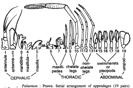
Instructions for dissection of Palaemon Study and draw the following appendage (19 pairs).
Cephalic appendages of Palaemon (Prawn)
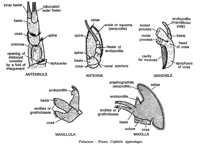
- Antennule: It is sensory and tactile in function and contains inner and outer feelers, basis, coxa, precoxa and stylocerite. Precoxa contains statocyst and is large. The basis is longer than coxa and carries 2 long sensory feelers. Outer feeler is divided into 2 unequal branches.
- Antenna : It is sensory, excretory and balancing, and it consists of coxa and basis which bears an expanded leaf-like exopodite, called as squama, and a long narrow feeler.
- Mandible : Mandibles are short and stout, lying one on each side of the mouth. It is masticatory in function and consists of coxa. The coxa is densely calcified to form powerful jaws. Its proximal part is spoon-shaped, having cavity for muscle insertion and is called as apophysis, while the distal part is called as head. Head contains stout molar process and a flat plate-like incisor process. Its outer border also contains a 3-jointed mandibular palp.
- Maxillula : It also helps in manipulation of food and consists of coxa, basis, gnathobases and endopodite.
- Maxilla: This manipulates food and is composed of coxa, basis, gnathobases and endopodite. The exopodite is large and forms a fan-shaped structure, called scaphognathite. It is also respiratory in function.
Thoracic appendages of Palaemon (Prawn)
It comprises of anterior 3 pairs of maxillipedes and posterior 5 pairs of walking legs.
- First maxillipede :
- It is formed by flattened, leaf-like coxa and basis
- Coxa contains a bilobed leaf-like epipodite
- Endopodite is small and unsegmented.
- The exopodite is also unsegmented but elongated, with a basal plate-like expansion.
- Second maxillipede
- It comprises of coxa and basis. It is not so flattened
- Coxa is short and contains a small epipodite and gill on outer side and setae on inner side
- Basis is jointed to ischium of endopodite.
- Endopodite comprises of 5 segments namely ischium, merus, carpus propodus and dactylus.
- Exopodite is elongated and contains setae
- Third maxillipede :
- It is leg-like and comprises of coxa and basis
- Coxa has a small epipodite on its outer side and basis bears a slender unjointed exopodite and an elongated endopodite.
- The endopodite is composed of 3 podomeres, proximal podomere fused with merus, middle carpus and terminal one fused with dactylus.
- Setae are found all along exopodite and on inner margin of endopodite.
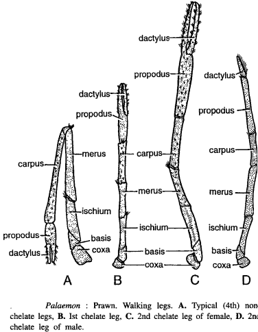
- Typical leg
- There are 5 pairs of walking legs differing from maxillipedes in the absence of exopodite and endopodite and also greater in size,
- A typical leg or 4th leg consists of 2-jointed protopodite and a 5-jointed endopodite.
- All the seven podomeres namely coxa, basis, ischium, merus, carpus, propodus and dactylus, are arranged in linear series and hinged together.
- First chelate leg
- In this, the propodus is prolonged beyond its articulation with the dactylus, so that 2 podomeres work one against the other like forceps blades forming chela or pincer. These legs are called as cheliped or chelate legs,
- Setae cover the entire surface.
- Second chelate leg
- All podomeres are considerable elongated
- In males, it is more powerful than in female. Third, fourth, and fifth pairs of legs are non-chelate and typical. In males, each fifth leg bears a male genital aperture on the arthrodial membrane between leg and thorax. In females, the genital aperture is found on the inner side of the coxa of third leg.
Abdominal appendages of Palaemon (Prawn)
There are six pairs of abdominal appendage. These are typical biramous appendages, one pair in each abdominal segment. They help in swimming and hence called as swimmerets or pleopods.
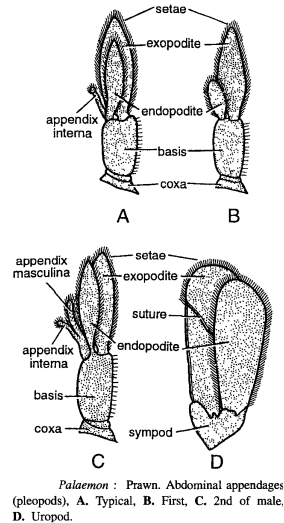
- Typical abdominal appendages (3rd appendages)
- The protopodite has ring-like coxa and a cylindrical basis,
- The basis bears flattened leaf-like smaller endopodite and a larger exopodite.
- From inner basal margin of endopodite, a small knob-headed rod-like structure arises, called as appendix interna.
- In female, the appendix interna of opposite appendages articulate with each other forming bridges to carry the eggs,
- The outer surface of basis and margins of exopodite and endopodite contain several setae,
- Remaining appendages slightly differ from typical ones.
- First abdominal appendage : Appendix interna absent and endopodite greatly reduced.
- Second abdominal appendage : In males only, in addition to appendix interna, there is additional rod-like and setae-bearing process called as appendix masculina, found between appendix interna and endopodite.
- Uropods : Sixth pair of appendages are called uropods lying one on either side of telson. In each uropod, coxa and basis are fused together to form a triangular sympod. Endo and exopodites are oar-like and their margins bear setae. Exopodite is bigger than endopodite and incompletely divided in the middle by a transverse suture.
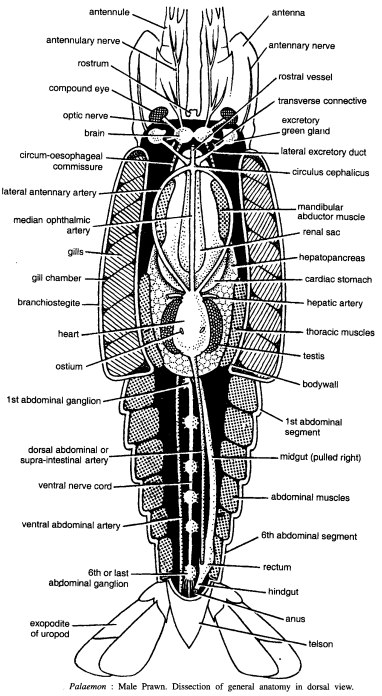
General anatomy of Palaemon (Prawn) Procedure
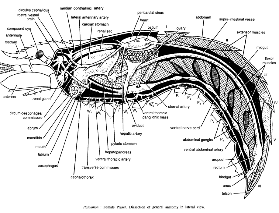
For Dissection of Palaemon (Prawn)take the specimen in hand or dish and lift carapace from lateral side. Cut loose the carapace at its anterior margin to remove it completely. Remove all the abdominal terga and pleura with scalpel and forceps and expose the abdominal muscles. Pin the specimen in dissecting dish and study the various organs.
Procedure for dissection of palaemon Reproductive organs : To expose the gonads fully, remove the heart and two narrow longitudinal extensor muscles.
Procedure for dissection of palaemon (Prawn) Digestive system : Remove completely the gonads to expose digestive system. The stomach is found just beneath carapace, embedded in large digestive gland. The intestine is a narrow tube present in the groove of abdominal muscles. For hastate plate or gastric mill, cut the stomach at both ends and make a dorsal incision on the dorsal side. Spread it flat, clean, wash and then see its teeth and comb etc. with the lens.
Procedure for dissection of palaemon (Prawn) Nervous system: For nervous system first expose the nerve cord in the abdominal region. For this, cut medianly with the scalpel between the large flexor muscles. Press these muscles and pin them in the dissecting tray. As the muscles are stretched, the nerve cord is very clearly seen with ganglionic swellings. Proceed from posterior side, cutting the middle line with the scissors through the chitinous endophragmal skeletal plates found on the ventral side of the thorax and exposing underlying nerves till brain is exposed. Study the various parts.
Musculature : It occupies greater part of the body segmentally arranged and inserted on the inner surface of the cuticle and its internal foldings called apodemes. The cut muscles of the abdomen are very clear, (i) Extensor muscles for straightening abdomen, (ii) Flexor muscles for bending abdomen, (iii) Muscles for moving appendages and for alimentation are also found.
Digestive system of Palaemon (Prawn)
It is well developed with associated glands and is divided into three regions: Foregut, midgut and hindgut. The foregut consists of the following parts
- Mouth : It is a large aperture on the ventral side of the head bounded by labrum, mandible and labium.
- Buccal cavity : Mouth leads into a short buccal cavity.
- Oesophagus: It is a short duct arising from buccal cavity and it communicates with stomach. It extends upwards.
- Stomach : It is a wide chamber consisting of cardiac and pyloric parts. Ventrally stomach is surrounded by orange-red hepatopancreas. Internally cardiac stomach shows cuticular thickening called as hastate plate. The midgut is a very short and slender duct, which ascends between hepato-pancreas to extend backwards. The hind gut extends from midgut up to anus. The terminal part is modified as rectum.
Circulatory or vascular system : If consists of pericardium heart and blood vessels. Arteries are ophthalmic, antennary, hepatic, sternal, supra-intestinal, thoracic and ventro-abdominal. Excretory organs : Renal glands. Nervous system: It consists of well-developed nerve cord and brain.
Other structures
- Parts of appendages, uropods, telson, eyes, gills (pleurobranchial), arthrobranchial and podobranchial) and gonads.
- Various structures seen in section of lateral view are rostrum, antennule, compound eye, brain, rostral vessel, circulus cephalicus, median ophthalmic artery, lateral antennary artery, cardiac stomach, renal sac, pericardial nerve, heart, ostium, ovary, abdomen, supra-intestinal vessel, extensor muscle, midgut, flexor muscle, telson, anus, hind gut, rectum, uropod, ventral abdominal artery, abdominal ganglia, ventral nerve cord, sternal artery, ventral thoracic ganglionic mass, oviduct, hepatic artery, pyloric stomach, hepatopancreas, ventral thoracic artery, transverse commissure, cephalothorax, oesophagus, labium, mouth, mandible, labruus, circum-oesophageal commissure, renal gland and antenna.
- Various structures seen in dissection of general anatomy in dorsal view are antenna, antennary nerve, rostral vessel, transverse connective excretory duct, circulus cephalicus, mandibular adductor muscle, renal sac, hepatopancreas, cardiac stomach, hepatic artery, thoracic muscles, testis, body wall, 1st abdominal segment, midgut, abdominal muscle, 6th terminal segment, rectum, hind gut, anus, telson, exopodite of uropod, last abdominal ganglia, ventral abdominal artery, dorsal supra-intestinal artery, 1st abdominal ganglia, ostium, heart, branchiostegite, gill chamber, gills, median ophthalmic artery, lateral antennary artery, circum-oesophageal commissure, brain, optic nerve, compound, eye, rostrum antennary nerve and antennule.
Nervous system of Palaemon (Prawn)
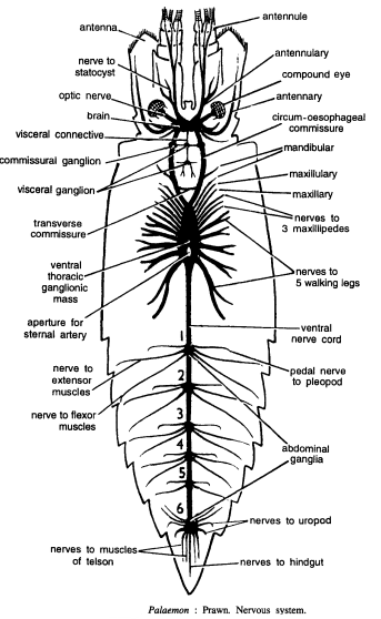
The nervous system of Palaemon is the most favourite dissection of the students. It comprises of :
- Brain or supra-oesophageal ganglia : It is a bilobed ganglion, situated at the base of rostrum. On each side, brain gives antennulary nerve to antennule, optic nerve to compound eye, statocystic nerve to statocyst, antennary nerve to antenna and tegumental nerve to labrum
- Circum-oesophageal commissures : Brain gives rise to a pair of thick posterior circum-oesophageal commissures, which surround oesophagus and unite together ventrally with sub-oesophageal ganglion. A transverse connective connects the two commissures. The sub-oesophageal ganglion is fused with anterior part of ventral thoracic ganglionic mass.
- The ventral thoracic ganglionic mass : It is formed by the fusion of eleven segment a cephalothoracic ganglia, which consequently gives 11 ‘pairs of nerves’. The first three pairs innervate the mandibles, maxillulae and maxillae; the next 3 pairs supply to 3 pairs of maxillipedes and the remaining 5 pairs of nerves supply to walking legs.
- The ventral nerve cord: It runs in the abdomen, fOlming 6 ganglionic masses, from where nerves are given out to pleopods, musculature and uropods.
Share this:

Discover more from ZOOLOGYTALKS
Subscribe to get the latest posts sent to your email.
Individuals trying to find low-priced Car Insurance
in Las Vegas Nevada usually begin through comparing quotes
online. You may spare money on Car Insurance in Las Vegas Nevada through sustaining a clean driving
report and choosing a much higher insurance deductible. Some firms providing Car Insurance in Las Vegas Nevada also offer loyalty markdowns for long-lasting customers.
Be sure to talk to about all available markdowns when purchasing Car
Insurance in Las Vegas Nevada.