
INTRODUCTION
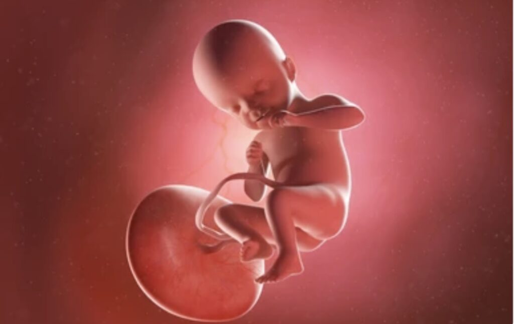
Placenta and types of placenta: The placenta is a vital organ in most mammals, including humans, that serves as a connection between the developing fetus and the mother. It facilitates the exchange of nutrients, gases, and waste products between the maternal and fetal circulatory systems, providing essential support for fetal growth and development. While the basic function of the placenta remains consistent across mammalian species, there are various types of placentation, each with unique structural and functional characteristics. The placenta is a remarkable organ essential for the development and sustenance of a fetus during pregnancy. Understanding its structure, function, and types is crucial in appreciating its pivotal role in pregnancy.
STRUCTURE OF PLACENTA
The placenta typically develops from the chorion, one of the outer membranes that surround the embryo. It consists of maternal and fetal components intricately intertwined to facilitate exchange and support:

Maternal Side
Derived from the endometrium (uterine lining), the maternal side of the placenta is characterized by decidual tissue and maternal blood vessels. These vessels undergo modifications to form uteroplacental arteries and veins, allowing for blood flow to and from the placenta.
Fetal Side
The fetal side of the placenta develops from the chorionic sac and contains fetal blood vessels immersed in chorionic villi. These villi are finger-like projections that extend into pools of maternal blood, maximizing surface area for efficient exchange of nutrients and waste products.
FUNCTIONS OF PLACENTA

The placenta serves several vital functions crucial for fetal development and maternal health:
- Nutrient and Gas Exchange: Through the chorionic villi, the placenta facilitates the transfer of oxygen, nutrients (such as glucose and amino acids), and antibodies from the mother’s bloodstream to the fetus. Simultaneously, carbon dioxide and waste products are transported from the fetus to the mother’s circulation for elimination.
- Endocrine Function: The placenta produces hormones like human chorionic gonadotropin (hCG), estrogen, and progesterone, which are essential for maintaining pregnancy, preparing the breasts for lactation, and supporting fetal growth and development.
- Immunological Barrier: It acts as a barrier against pathogens, preventing most microorganisms from crossing from the maternal to the fetal circulation and protecting the developing fetus from infections.
- Waste Elimination: The placenta removes fetal metabolic wastes, such as urea and uric acid, which are then eliminated through the maternal kidneys.
TYPES OF PLACENTA
Understanding the different types of placenta is essential for comprehending the diverse reproductive strategies and adaptations seen in mammals.
Epitheliochorial Placenta
Epitheliochorial placentation is found in some species of ungulates, including cows, sheep, and pigs. In this type of placenta, there are multiple layers of tissue separating the maternal blood supply from the fetal chorion, including the maternal endometrium and fetal chorionic epithelium. This arrangement provides significant protection for the developing fetus but limits the efficiency of nutrient exchange compared to other types of placentation.
Endotheliochorial Placenta
Endotheliochorial placentation is characteristic of carnivores such as dogs, cats, and bears. In this type of placenta, the maternal uterine endothelium is in direct contact with the fetal chorion, with only the maternal capillaries separating the two layers. While this arrangement improves nutrient exchange compared to epitheliochorial placentation, it still involves multiple tissue layers, which can restrict the efficiency of gas exchange.
Hemochorial Placenta
Hemochorial placentation is the most efficient type of placenta in terms of nutrient and gas exchange and is found in primates, including humans, as well as rodents and some insectivores. In this type of placenta, the maternal blood comes into direct contact with the fetal chorion, with no intervening tissue layers. This direct contact allows for highly efficient exchange of nutrients, gases, and waste products between the maternal and fetal circulatory systems, facilitating the rapid growth and development of the fetus.
Syndesmochorial Placenta
Syndesmochorial placentation is found in some species of rodents and insectivores. In this type of placenta, the fetal chorion comes into direct contact with maternal tissues, but there is some degree of fusion between the maternal and fetal epithelia, forming specialized areas known as areolae. These areolae allow for increased surface area and improved nutrient exchange compared to endotheliochorial placentation but still involve some degree of separation between the maternal and fetal circulatory systems.
Endotheliochorial-Hemomonochorial Transition Placenta
This type of placenta is unique to some species of bats. In early gestation, the placenta is endotheliochorial, similar to carnivores, with the maternal endothelium in direct contact with the fetal chorion. However, as gestation progresses, the placenta undergoes a transition to a hemomonochorial arrangement, with the maternal blood coming into direct contact with the fetal chorion. This transition allows for improved nutrient exchange as gestation advances and metabolic demands increase.
Hemomonochorial Placenta
Hemomonochorial placentation is found in some species of rodents and insectivores, including guinea pigs and shrews. In this type of placenta, the maternal blood comes into direct contact with the fetal chorion, similar to hemochorial placentation. However, unlike true hemochorial placentas, which have a single layer of fetal trophoblast cells separating the maternal and fetal circulatory systems, hemomonochorial placentas have multiple layers of fetal trophoblast cells, providing additional structural support.
CONCLUSION
In summary, the diversity of placental structures reflects the diverse reproductive strategies and adaptations seen in mammals. From the multi-layered epitheliochorial placenta of ungulates to the highly efficient hemochorial placenta of primates, each type of placentation represents a unique solution to the challenges of supporting fetal development within the maternal environment. Understanding the differences between these types of placenta is essential for appreciating the complexity of mammalian reproduction and evolution.
Share this:

Discover more from ZOOLOGYTALKS
Subscribe to get the latest posts sent to your email.